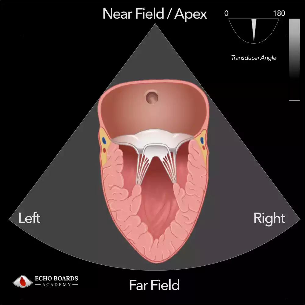
The sector display for a TEE machine is a pie-shaped image that shows the structures of the heart and surrounding tissues. The top of the sector represents the tissue closest to the TEE probe, while the bottom of the sector represents the tissue farthest away.
The width of the sector is determined by the field of view (FOV) setting on the TEE machine.
The sector display is the most common type of display used in TEE imaging. It is useful for visualizing the overall structure of the heart, as well as for specific views such as the long-axis view, short-axis view, and bicaval view.
Understanding how the display is organized is pivotal to:
- Determine the depth of field
- Adjust gain settings
- Optimize the width of the sector
By mastering these settings, you can tailor the image quality to suit the clinical question at hand.
Centering the Image
It is important to center a heart structure in the sector display when performing TEE for the following reasons:
- To obtain the best possible image quality. When the heart structure is centered in the sector display, it is more likely to be in focus and well-lit. This is because the TEE probe is able to send and receive sound waves more effectively when the heart structure is centered in the image.
- Allows for proper rotation of omniplane transducer around the heart structure of interest.
- To avoid artifacts. Artifacts are distortions in the TEE image that can be caused by a variety of factors, such as the patient’s anatomy, the TEE probe position, and the machine settings. By centering the heart structure in the sector display, you can help to minimize the risk of artifacts.
- To make it easier to measure and assess the heart structure. When the heart structure is centered in the sector display, it is easier to measure its size, shape, and function. This is because the TEE machine is able to calculate these measurements more accurately when the heart structure of interest is in the center of the image.
In addition to these reasons, centering the heart structure in the sector display also makes it easier to navigate and to obtain different views of the heart.
If you are having difficulty centering the area of interest in the sector display, you can ask a more experienced colleague for assistance.
Key Points When Using The Sector Display for TEE.
- The sector display is a two-dimensional image, so it does not show the depth of structures. To get a better understanding of the depth of structures, you can use 3D TEE imaging or color Doppler imaging.
- The sector display can be affected by the TEE probe position and orientation. It is important to position the probe correctly and to adjust the orientation as needed to obtain the best possible image.
- The sector display can also be affected by the patient’s anatomy. For example, patients with a dilated ascending aorta may have difficulty obtaining a the standard image angles.
Overall, the sector display is a versatile and informative tool for TEE imaging. It can be used to visualize the overall structure of the heart, as well as specific views that are needed to assess different aspects of cardiac function.
Tips for Using the Sector Display For TEE
- Use the FOV setting to adjust the width of the sector. A narrower FOV will provide a more detailed image of the structures in the center of the sector, while a wider FOV will provide a more general view of the heart.
- Use the zoom function to magnify the image. This can be helpful for visualizing small structures or for getting a more detailed view of a specific area.
- Use the depth setting to adjust the depth of the image. This can be helpful for visualizing structures that are farther away from the probe.
- Use the image orientation controls to rotate and flip the image. This can be helpful for obtaining the best possible view of the heart.
If you are new to TEE imaging, it is important to practice using the sector display to obtain different views of the heart.
Conclusion
Accurate echocardiographic imaging is a multifaceted process that relies heavily on the expertise of the healthcare provider. By understanding and utilizing the different imaging planes, probe movements, and display modes, clinicians can significantly enhance the diagnostic utility of the examination.